Abstract: Deep-learning-based Segmentation of Ellipsoid Zone And Geographic Atrophy On Real-World Optical Coherence Tomography
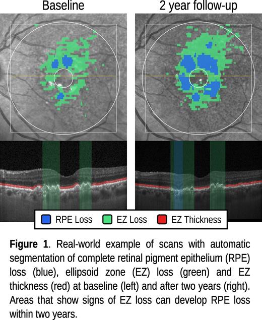
Method to use AI to automatically segment in optical coherence tomography (OCT) images of the retina where photoreceptor or retinal pigment epithelium cells are lost. This allows to quantify the extent of geographic atrophy secondary to age related macular degeneration in the retina.
Wolf-Dieter VoglOliver LeingangHlynur SkulasonAmir SadeghipourAriadne Whitby
Investigative Ophthalmology & Visual Science June 2025, Vol.66, 5848. doi:
Purpose: Geographic Atrophy (GA) study data often have strict inclusion criteria, such as lesion size, which can limit the diversity of training examples for AI-based deep learning models. This restriction may result in biased validation outcomes and diminished performance when applied to real-world data. To address this, we trained novel deep learning models on a combined study and real-world dataset to segment relevant GA biomarker, complete retinal pigment epithelium (RPE) loss, ellipsoid zone (EZ) thickness, and EZ loss. The models were validated on an independent real-world dataset to evaluate their robustness and generalizability.
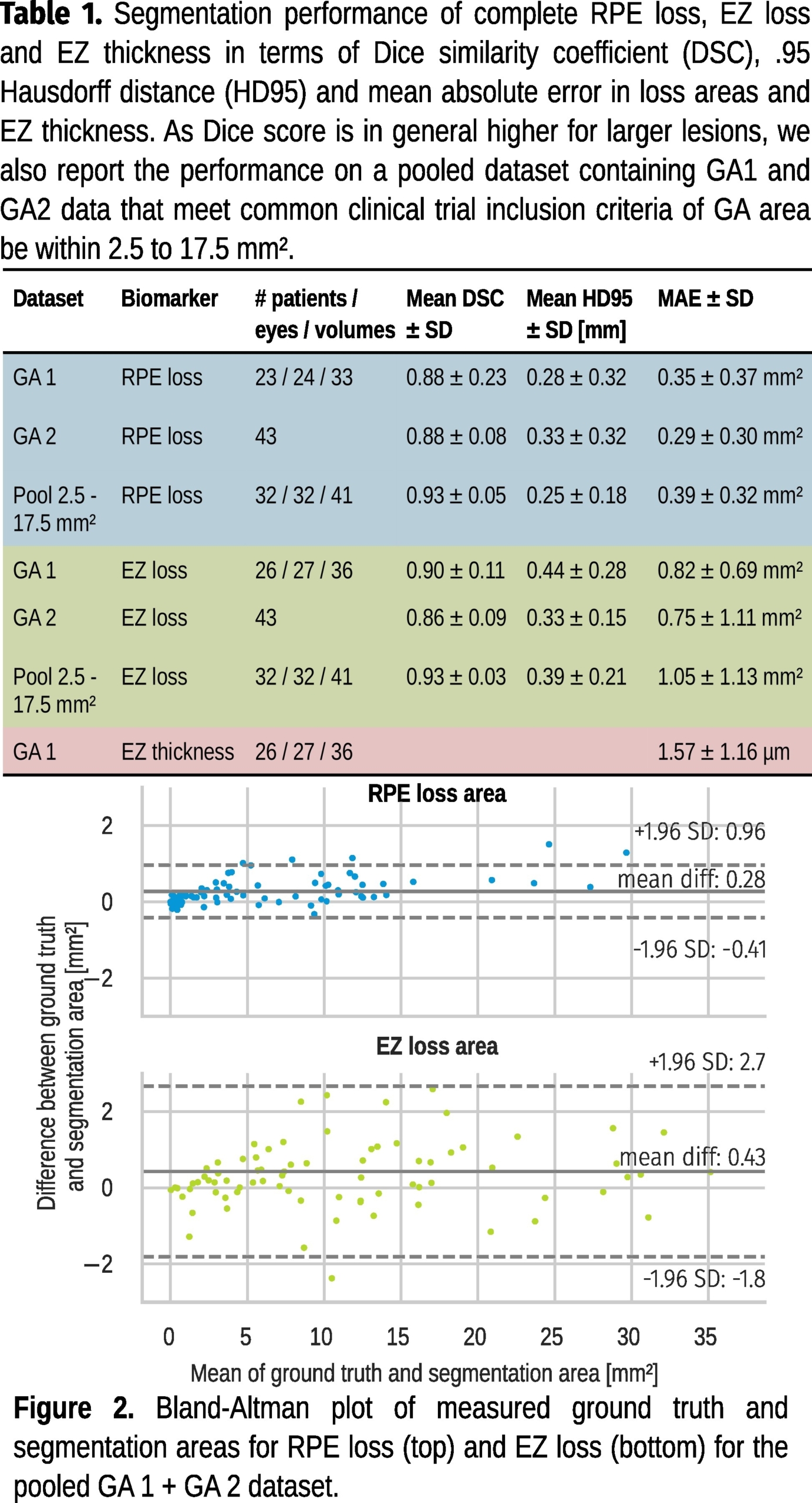
Methods: Experienced readers annotated areas of complete RPE loss, the inner boundary of the EZ (IB-EZ) layer, interruptions to the EZ layer, and the outer boundary of the outer photoreceptor layer (OB-OPR) on SD-OCT images from a Heidelberg Spectralis device according to a rigorous annotation protocol. A novel 3D-to-2D convolutional neural network (CNN) was trained to segment RPE loss. A 2D CNN network was trained to segment the region between the IB-EZ and OB-OPR. The performance of these models was evaluated on a hold-out test set from the training dataset (GA1) and on a real-world dataset (GA2).
Results: For model training a total of 298 Heidelberg Engineering Spectralis SD-OCT volumes were selected from a clinical trial (199 volumes) and clinical practice(99 volumes). Eyes showed conditions of GA (222), neovascular AMD (17), intermediate AMD (40) and no pathology (19). Performance was evaluated on a validation set of 43 volumes (GA2), randomly selected from a real-world pool of 950 eyes with GA, stratified by RPE atrophy size (median: 1.86mm2, IQR: 6.74). Mean Dice ± SD of RPE loss segmentation for GA1 and GA2 was 0.88±0.23 and 0.88±0.08, respectively. For EZ loss we report a Dice of 0.90±0.11 and 0.86±0.09 on GA1 and GA2, respectively. Considering study inclusion criteria GA lesion size of 2.5 to 17.5mm2 only, RPE loss and EZ loss Dice is 0.93±0.05 and 0.93±0.03, respectively, for pooled GA1+GA2.
Conclusions: By utilizing novel deep learning models trained on a diversity of study and real-world datasets, we can robustly and precisely segment and measure RPE loss, EZ loss, and EZ thickness. Accurate biomarker measurement is essential for early detection of GA, monitoring its progression, and evaluating the efficacy of therapeutic interventions.
This abstract was presented at the 2025 ARVO Annual Meeting, held in Salt Lake City, Utah, May 4-8, 2025.